
Ophthalmology
Cataract Surgery
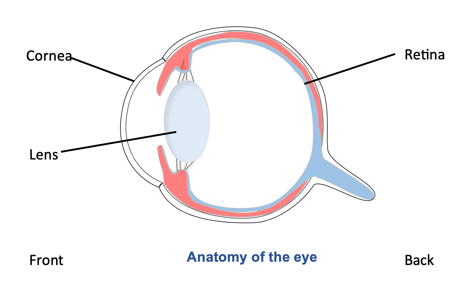
Phacoemulsification
A cataract is a change to the proteins that make up the lens, resulting in an opacity which affects vision. Cataract surgery is a commonly performed surgery in dogs, and the aim is to improve vision and improve quality of life. There are possible complications, and the risk of these is higher than in human patients.
The surgery:
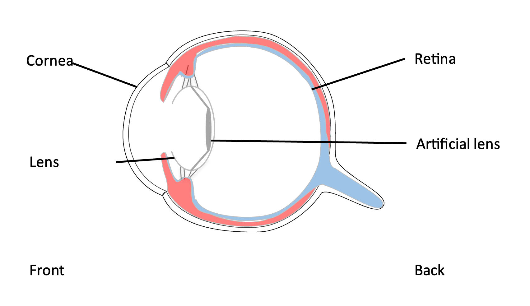
The surgery is similar to that performed in people and uses a process called phacoemulsification. Phacoemulsification is the use of ultrasound energy to break up the lens material and remove it. Once the cataract material is removed, an artificial lens is usually placed inside the lens capsule (the stretchy bag that holds the lens material), to restore focus. Surgery is performed under general anaesthesia. We also use a drug that relaxes the muscles in the body, keeping the eye central and relaxed. During this process patients need to be kept on a ventilator, and closely monitored by our experienced team.
Success rates for cataract surgery:
Surgical success in dogs depends on several factors. This includes species (e.g. dog or cat), breed (some breeds are predisposed to complications such as glaucoma), pre-existing inflammation inside the eye (uveitis) and medical complications, such as diabetes. The most reported statistic across several studies of cataract surgery in dogs is that around 90% of eyes are visual one year after surgery. It is a little higher for cats.
Risks of cataract surgery:
Anaesthesia carries some risk, but this can be minimised by careful patient choice and pre-operative assessment. All surgery carries some risk; the specific risks associated with cataract surgery are the development of secondary glaucoma (high pressure in the eye) and retinal detachment. These complications can be blinding or cause ocular discomfort; and additional treatment may be required.
Possible complications that can occur after cataract surgery and signs to look out for
Inflammation within the eye is normal after cataract surgery and will be closely monitored during at post-operative examinations. Corneal ulcers (a problem with the surface of the eye) can occur after cataract surgery and should be dealt with immediately.
The main signs of any complication after ocular surgery are increased discomfort – squinting and/or tearing – discharge from the eye, and redness of the ‘white of the eye’. If you have any concerns about your pet’s eyes after the operation, you should contact us immediately as some of the complications, such as glaucoma, can rapidly lead to blindness. It is possible that complications can arise even years after surgery, and we monitor patients after cataract surgery for the rest of their lives.
Pre-operative tests
Most patients have surgery when their cataracts are quite advanced. These means it can be difficult or impossible to see to the back of the eye. In order to make sure the back of the eye is healthy, and the patient will benefit from surgery, we perform:
Ocular Ultrasound
This enables the ophthalmologist to check that the retina is in place and there are no abnormalities that may complicate surgery. Ultrasound is usually performed on conscious patients, with local anaesthetic drops to numb the surface of the eye.
Electroretinography (ERG)
An electroretinogram (ERG) is a test to assess the function of the retina, the light sensitive tissue at the back of the eye. Retinal disease is common in dogs and, if present, may mean that the patient won’t benefit from surgery. We usually perform this test immediately before surgery, under anaesthesia.
Blood tests
We usually perform a blood test to provide some information about general health, to ensure your pet is safe to undergo anaesthesia.
Cardiac ultrasound (echocardiogram)
If any abnormalities are heard on listening to your pet’s heart, we may recommend an assessment by a cardiologist prior to anaesthesia.
Dental treatment
Dental disease can increase the likelihood of post-operative infection and inflammation in the eye. Therefore, we may recommend dental treatment with your primary vet before planning cataract surgery.
Diabetic dogs – special considerations
Ideally, diabetes should be well-controlled before surgery. However, diabetic cataracts develop rapidly and are associated with inflammation in the eyes, making surgery relatively urgent. In practice, surgery is often performed before diabetic control is perfect. However, it may not be appropriate to risk surgery in an unstable patient. Diabetic controlled can also be disrupted by anaesthesia, so you can expect some increase in thirst and frequency of urination immediately after surgery.
Hospital stay
Most pets stay for 2 nights, the night before and the night following surgery, as they require intensive eye medications, pain relief and monitoring, (e.g. regular measurement of the pressure inside the eye). However, we treat each pet as an individual, and rarely it may be preferable for your pet to return home on the day of surgery. If this is the case you will be asked to return in the morning with them, so they can be assessed.
Vision after surgery
Your pet will be able to see after surgery, but vision may be blurry at first due to inflammation inside the eye after the surgery. As the inflammation subsides the vision will improve. If an artificial lens is placed, then vision will be restored quickly. Sometimes we are unable to place a lens, but for these cases functional vision is still restored (the patient can see well enough to navigate around objects), but close vision will be less precise. Over time the brain will adapt and vision will continue to improve.
Aftercare
- The first few weeks will require an intensive regime of eye medications, and they will need to be kept calm. There may be 5 or even 6 different eye drops, up to four times a day.
- An Elizabethan collar will be worn at all times for the first 14 days.
- Short, gentle harness walk only, to prevent excess pressure around the neck, for 2-3 weeks. No jumping onto furniture or running up stairs. After 3 weeks gentle exercise can slowly be resumed.
- There will also be oral medication for the first few weeks.
- We examine patients at 1 week following surgery, 2 weeks after that, 4-6 weeks after that, 3 months later and then every 6-12 months, (if all goes well) for the rest of their lives. You may need to return for frequent visits in the first year if there are any complications.
Cost of surgery:
| One eye | £4750 |
| Both eyes (at the same time) | £6000 |
What is included:
These prices include pre-operative tests (blood sampling, ocular ultrasound, electroretinogram recording), medication for immediate use after surgery and three follow-up examinations.
What is not included:
Ongoing medication. Many of the eye drops used are human eye drops, and we can often write prescriptions so that you can buy these from a ‘human’ pharmacy, where they are cheapest. Please ask us about this if we do not mention it.
Follow-up after the first three examinations. These appointments are charged at the time of examination and are £210.
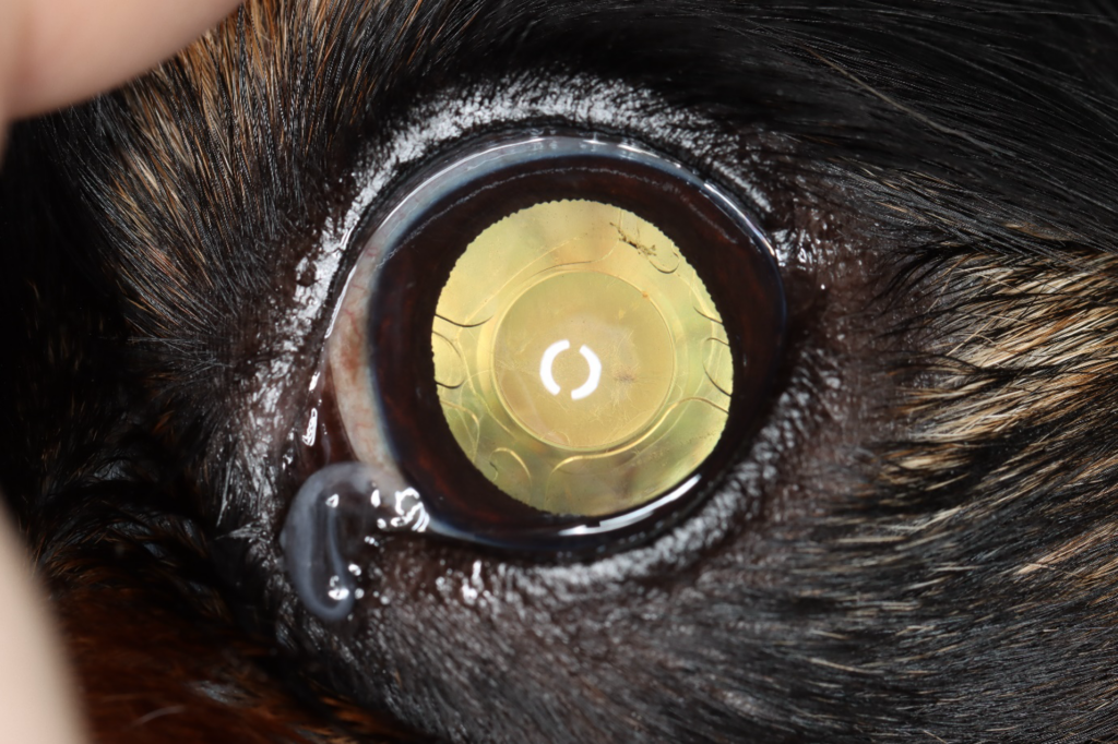
This photograph shows an eye with an artificial lens placed, after cataract surgery.