
A vet-to-vet piece from South East Vet Referrals.
Once a tumour is confirmed or strongly suspected, the next question is rarely only what it is. It is how far it has gone. Two patients with the same tumour type can need very different plans, and what usually separates them is stage.
That is why our oncology service offers staging as a defined step. An oncology assessment, then imaging chosen to fit the case, using radiographs, ultrasound or CT as needed, to look at the extent of the primary tumour and to look for metastasis. Where imaging has already been done during the diagnosis, it can be used, and the plan can start from there.
Stage and grade answer different questions
The two terms often travel together, and they are easy to blur. They describe different things.
Staging asks how far the disease extends. It looks at three things: the primary tumour locally, the regional lymph nodes, and distant metastasis. This is the familiar TNM framework.
Grade describes the primary tumour itself, from its histological appearance. It comes from histopathology rather than imaging, and it tells you about the tumour’s likely behaviour rather than where it has already gone.
| Stage | Grade | |
|---|---|---|
| The question | How far has it extended? | How does the primary tumour look under the microscope? |
| Where it comes from | Assessment, imaging and sampling | Histopathology of the primary tumour |
| What it looks at | Local extent, lymph nodes, distant metastasis | The primary tumour only |
The two work together. For some tumour types, grade can make spread considerably more or less likely. Staging then looks for whether it has happened. A high grade on a histopathology report is often the moment staging moves up the list.
Why stage before deciding on treatment
Staging can shape almost every decision that follows.
What treatment is needed. Local disease and disseminated disease may call for quite different approaches, so knowing the extent early helps avoid committing to a plan that does not fit.
Planning surgery. The extent of the primary tumour, and whether nodes are involved, can inform what surgery should aim to achieve, or whether surgery is the right first step at all.
What comes after surgery. Staging can help decide whether post-surgical chemotherapy may be needed. Where radiation may belong in the plan, the oncology team can advise on that early and coordinate onward referral, so the right cases reach the right centre.
Prognosis. Stage is one of the most useful things to have in hand for the owner conversation. It helps set realistic expectations, in both directions, before decisions are made.
How likely is spread? Some published figures
Metastatic behaviour varies widely between tumour types, and within a type it can vary with grade. That is one reason a staging plan is not one size fits all. The figures below are approximate published rates for some common tumours, drawn from the 2026 AAHA Oncology Guidelines for Dogs and Cats.
| Tumour | Reported metastatic behaviour |
|---|---|
| Canine osteosarcoma | Pulmonary micrometastases reported in over 90% at presentation. Lung is the most frequent site; bone, kidney, liver and heart are also reported. Skeletal metastases are less common. |
| Canine splenic haemangiosarcoma | Metastatic rate approaches 100%. Liver is the most commonly reported site. |
| Canine oral malignant melanoma | Approximately 80%, commonly to regional lymph nodes and then the lungs. |
| Canine soft tissue sarcoma | Approximately 20% overall, rising with grade: around 15% for grades 1 and 2, and around 50% for grade 3. Clinically apparent metastases may develop relatively late. |
| Canine mammary carcinoma | Under 50% of malignant tumours overall. Risk increases with adverse features, including larger size, high grade and lymphovascular invasion. |
| Canine mast cell tumour | Strongly grade dependent (Patnaik grading): rare for grade 1, approximately 20% for grade 2, and 50 to 100% for grade 3. |
| Feline mammary carcinoma | Highly metastatic, approximately 80 to 90%, commonly involving lymph nodes, liver and lungs. |
| Feline soft tissue sarcoma | Under 10% for non-injection-site, low-grade tumours; over 25% for high-grade and/or injection-site sarcomas. |
| Feline squamous cell carcinoma | Overall metastatic rate described as low. Oral SCC is locally aggressive; cutaneous SCC is often more slowly progressive. |
These are population figures. They cannot predict what an individual patient will show, which is exactly why staging the patient in front of you is worth doing. Where metastases may develop late, follow-up imaging can be built into the plan.
A case: Barney
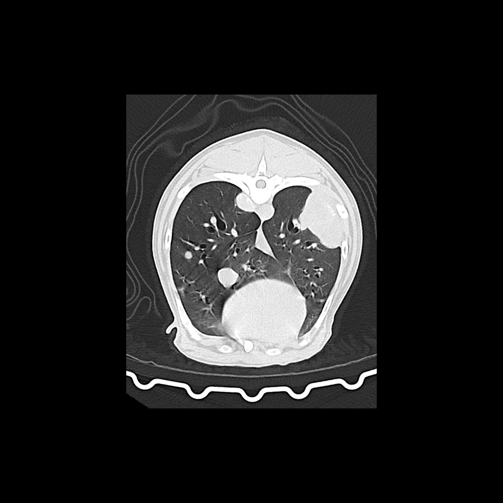
Barney, a nine-year-old male neutered Labrador, had a tumour removed from a toe earlier in the year. Chest radiographs were clear, but histology was suspicious for osteosarcoma.
Osteosarcoma has a very high rate of spread to the lungs. Even where metastases are not seen at diagnosis, they are usually present microscopically, and small metastases may not be visible on thoracic radiographs. CT can show smaller lesions.
Several months later Barney came to us with lameness in several limbs. Joint taps led to a diagnosis of immune-mediated polyarthritis, which was likely paraneoplastic. CT showed masses throughout the lungs, suggestive of metastases from the original toe tumour. Sadly, Barney was put to sleep.
The case is a reminder of why the choice of staging imaging matters for tumour types with a known high metastatic rate, and why a clear first result is worth revisiting when new signs appear.
What staging can involve at SEVR
Oncology assessment. Most cases start with a phone or video consultation, and an in-person consultation at Sevenoaks can be arranged if the family prefers.
Imaging chosen to the case. Chest radiographs, abdominal ultrasound or CT, with sampling where it is needed. Not every patient needs every modality, and the aim is the imaging that answers the question for that tumour. For tumours with a high rate of spread to the lungs, CT may pick up smaller lesions than radiographs can.
Lymph node mapping CT for more complex cases. CT lymphangiography can help identify the lymph nodes draining the tumour site, which can matter when nodal status would change the plan. It tends to be most useful where it is hard to tell which lymph nodes will be involved, especially in the head and neck.
What the day looks like. Staging is usually done as an outpatient, so most patients are with us for the day. It is carried out under sedation or anaesthesia, and most cases also include some form of sampling, either cytology or histology. Imaging is interpreted on site, so initial findings are available in real time. The full detailed CT report usually follows within around 24 to 48 hours, and cytology or histology results can take anywhere from a day to around a week.
Our staging packages are listed on the oncology fees page.
When a staging conversation is worth having
- A confirmed malignant tumour where the treatment plan depends on how far it extends
- Before surgery on a tumour type with known metastatic potential
- When histopathology comes back at a higher grade than expected
- When you are unsure which imaging would actually answer the question for a particular tumour
Plenty of practices already run some of this staging themselves, and that work is useful. A conversation can help decide what, if anything, to add, and the case can stay with you if that suits the patient and the owner.
The bottom line
Grade tells you about the tumour. Stage tells you about the patient. Both matter, and the treatment plan is usually stronger for having both before decisions are made.